Pre-Procedural Activities
10%of exam
Performing Resting ECG
14%of exam
Analyzing 12-Lead ECG
22%of exam
Performing Stress Tests
10%of exam
Ambulatory Monitoring
4%of exam
Performing Rhythm Analysis
40%of exam
Quick Facts
- Exam
- CCT
- Credential
- Cardiographic Technician
- Body
- CCI
- Items
- 130 (110 scored)
- Time
- 2 hours
- Pass
- 650/900 scaled
- Fee
- $175
- Level
- Entry-level
Pre-Procedural Steps
- Verify order
- Correct test ordered
- Two identifiers
- Name and DOB
- Consent
- Explain the procedure
- Skin prep
- Clean, dry, abrade
- Landmarks
- Palpate intercostal spaces
Limb Lead Colors
Snow over grass; smoke over fire
Artifact vs Arrhythmia
Artifact
- Normal underlying rhythm
- Patient looks stable
- Moves with motion
Arrhythmia
- True rhythm change
- Symptoms may occur
- Consistent abnormal complexes
Check pulse and patient
Normal Values & Grid
- PR interval
- Normal duration0.12-0.20s
- QRS duration
- Normal width<0.12s
- QT interval
- Rate-dependent limit<0.44s
- P wave
- Width and height limit<0.12s, <2.5mm
- Heart rate
- Normal sinus range60-100 bpm
- Small box
- 0.04s wide, 1mm0.04s / 0.1mV
- Large box
- 5 small boxes0.20s
- Paper speed
- Standard recording speed25 mm/s
- Calibration
- Standard gain10 mm/mV
- ST elevation
- Significant threshold≥1mm limb
Limb Lead Placement
- RA
- Right armWhite
- LA
- Left armBlack
- LL
- Left legRed
- RL
- Right leg groundGreen
- Lead I
- RA to LA
- Lead II
- RA to LL
- Lead III
- LA to LL
- aVR, aVF, aVL
- Augmented limb leads
- Einthoven's triangle
- I + III = II
Precordial Leads V1-V6
- V1
- 4th ICS, right sternalSeptal
- V2
- 4th ICS, left sternalSeptal
- V3
- Midway V2 and V4Anterior
- V4
- 5th ICS midclavicular lineAnterior
- V5
- Anterior axillary, V4 levelLateral
- V6
- Midaxillary line, V4 levelLateral
- V4R
- Right-sided; RV infarct
- V7-V9
- Posterior wall leads
ECG Artifacts
- Wandering baseline
- Respiration, loose electrodes
- 60-Hz interference
- AC electrical, fine spikes
- Somatic tremor
- Muscle movement, jagged
- Motion artifact
- Patient movement spikes
- Loose lead
- Sudden erratic tracing
Bundle Branch Blocks
WiLLiaM MaRRoW
RBBB vs LBBB
RBBB
- RSR' in V1
- Rabbit ears
- Wide S in V6
LBBB
- Broad R in V6
- QS or rS in V1
- Notched R wave
V1 up vs down
MI Localization by Leads
- ST↑ in II, III, aVF→Inferior MI(RCA)
- ST↑ in V1-V2→Septal MI(LAD)
- ST↑ in V3-V4→Anterior MI(LAD)
- ST↑ in I, aVL, V5-V6→Lateral MI(LCx)
- Tall R, ST↓ V1-V2→Posterior MI(Get V7-V9)
- Inferior MI + V4R↑→RV infarct(Preload dependent)
Waves, Segments & Intervals
- P wave
- Atrial depolarization
- QRS complex
- Ventricular depolarization
- T wave
- Ventricular repolarization
- U wave
- Late repolarization; hypokalemia
- PR interval
- Start P to QRS0.12-0.20s
- PR segment
- End P to QRS
- QRS duration
- Ventricular activation width<0.12s
- QT interval
- Depolarize plus repolarize
- ST segment
- J-point to T
- J point
- QRS meets ST
- Isoelectric line
- Flat baseline reference
- R-wave progression
- Grows V1 to V6
MI Territories
RCA inferior, LAD front, LCx side
STEMI vs Ischemia
STEMI
- ST elevation
- Acute injury pattern
- Reciprocal changes
Ischemia
- ST depression
- T-wave inversion
- No ST elevation
Elevation vs depression
Conduction System
- SA node
- Cardiac pacemaker60-100
- AV node
- Delays impulse40-60
- Bundle of His
- AV to bundle branches
- Bundle branches
- Right and left
- Purkinje fibers
- Ventricular spread20-40
- Pathway
- SA→AV→His→Purkinje
Axis Quadrants
Leads I and aVF both up = normal
Axis Determination
- Normal axis
- -30° to +90°
- LAD
- -30° to -90°
- RAD
- +90° to +180°
- Extreme axis
- -90° to -180°
- Lead I + aVF
- Both up = normal
- LAD pattern
- I up, aVF down
- RAD pattern
- I down, aVF up
MI Localization
- Inferior
- II, III, aVFRCA
- Septal
- V1-V2LAD
- Anterior
- V3-V4LAD
- Anteroseptal
- V1-V4LAD
- Lateral
- I, aVL, V5, V6LCx
- Posterior
- Tall R, V1-V2RCA/LCx
- RV infarct
- V4R ST elevationRCA
Ischemia, Infarction & Hypertrophy
- ST elevation
- Acute injury / STEMI
- ST depression
- Ischemia or reciprocal
- T inversion
- Ischemia marker
- Pathologic Q
- Prior infarct scar
- Hyperacute T
- Earliest STEMI sign
- LVH
- Sokolow-Lyon ≥35mmS-V1 + R-V5/6
- RBBB
- RSR' in V1≥0.12s
- LBBB
- Broad R in V6≥0.12s
Stress Test Protocols
- Bruce
- 3-min stages, speed+grade
- Modified Bruce
- Two warm-up stages
- Naughton
- 2-min, ~1 MET steps
- Target HR
- 85% × (220−age)
- Max HR
- 220 − age
- MET
- Metabolic workload unit
- Stop test
- ≥2mm ST depression
- BP drop
- Ischemia warning sign
Ambulatory Monitoring Types
- Holter
- Continuous 24-48 hours
- Event monitor
- Patient-activated symptoms
- Loop recorder
- Continuous loop memory
- Telemetry
- Inpatient real-time
- Patient diary
- Log symptoms + activity
Rate Sequence
300, 150, 100, 75, 60, 50
Mobitz I vs Mobitz II
Mobitz I
- PR lengthens each beat
- Then drops one QRS
- AV node level
- Usually benign
Mobitz II
- Constant PR interval
- Sudden dropped QRS
- Below AV node
- May need pacemaker
Lengthens vs sudden drop
Rhythm ID Logic
- Check R-R regularity first→Assess regularity(Step 1)
- Irregular, no P waves→AFib(Fibrillatory baseline)
- Sawtooth flutter waves→Atrial flutter(Atrial 250-350)
- Narrow, regular, 150-250→SVT(P wave hidden)
- Wide, regular, >100→VT(Emergency)
- Chaotic, no QRS→VFib(Defibrillate)
- Flatline tracing→Asystole(No shock)
- Upright P, 60-100→Sinus rhythm(Normal)
Rate Calculation Methods
- 300 method
- 300 / large boxes
- 1500 method
- 1500 / small boxes
- 6-second method
- QRS in 6s × 10
- Sequence
- 300-150-100-75-60-50
- Regular rhythm
- Use box methods
- Irregular rhythm
- Use 6-second method
- Normal rate
- 60-100 bpm
AFib vs Atrial Flutter
AFib
- Irregularly irregular rhythm
- No P waves
- Fibrillatory baseline
Atrial Flutter
- Sawtooth F waves
- Atrial 250-350 bpm
- Often regular ventricles
Chaotic vs sawtooth
AV Block Differentiation
- PR >0.20s, all conduct→1° AV block(Constant PR)
- PR lengthens then drops→Mobitz I(Wenckebach)
- Constant PR, sudden drop→Mobitz II(May need pacer)
- P and QRS unrelated→3° block(Complete block)
- Regular P, some blocked→2:1 block(Watch closely)
Sinus & Atrial Rhythms
- Sinus rhythm
- Upright P, 60-100 bpm
- Sinus brady
- Sinus, rate <60
- Sinus tachy
- Sinus, rate >100
- Sinus arrhythmia
- Rate varies with breathing
- PAC
- Early abnormal P wave
- AFib
- Irregularly irregular, no P
- Atrial flutter
- Sawtooth, atrial 250-350
- SVT/PSVT
- Narrow, regular, 150-250
- WAP
- Wandering, varying P shapes
- MAT
- ≥3 P shapes, >100
VT vs SVT
VT
- Wide QRS complex
- Rate 100-250
- AV dissociation
- Life-threatening
SVT
- Narrow QRS complex
- Rate 150-250
- P waves hidden
Wide vs narrow QRS
Ventricular & Junctional Rhythms
- PVC
- Wide, early, no P
- Junctional
- Inverted/absent P, 40-60
- VT
- ≥3 PVCs, rate >100
- VFib
- Chaotic, no organized QRS
- Asystole
- Flatline, no activity
- PEA
- Organized rhythm, no pulse
- Idioventricular
- Wide escape, 20-40
- Bigeminy
- Every other beat PVC
- Couplet
- Two consecutive PVCs
- Torsades
- Polymorphic VT, long QT
PAC vs PVC
PAC
- Early P wave
- Narrow QRS
- Non-compensatory pause
PVC
- No preceding P
- Wide bizarre QRS
- Compensatory pause
Narrow vs wide QRS
AV Blocks
- 1° AV block
- PR >0.20s, constant
- Mobitz I
- PR lengthens, drops QRS
- Mobitz II
- Constant PR, drops QRS
- 3° block
- P and QRS independent
- 2:1 block
- Every other P conducts
Common Traps
V1/V2 lead placement
4th ICS correct ≠ Not 2nd ICS
Mobitz I vs II
Type I PR lengthens ≠ Type II PR constant
AFib vs flutter
AFib has no pattern ≠ Flutter is sawtooth
Two meanings of LAD
LAD = coronary artery ≠ LAD = left-axis deviation
Rate calculation methods
300 / large boxes ≠ 1500 / small boxes
Sinus arrhythmia is normal
Normal breathing variant ≠ Not pathologic
Wide tachycardia
Assume VT until proven ≠ Not always SVT
Last Minute
- 1.Weights: Rhythm 40%, 12-lead 22%
- 2.PR normal 0.12-0.20s
- 3.QRS normal <0.12s
- 4.Rate: 300 / large boxes
- 5.V1: 4th ICS right sternal
- 6.Inferior MI = II, III, aVF
- 7.Anterior MI = V3-V4 (LAD)
- 8.Mobitz II drops without warning
- 9.AFib = irregularly irregular
- 10.Flutter = sawtooth, 250-350
- 11.VT = wide, regular, >100
- 12.Target HR = 85%×(220−age)
- 13.SA→AV→His→Purkinje pathway
- 14.Normal axis: I and aVF up
 Free CE
Third-party resources · We may earn a commission from purchases.
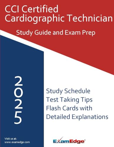
CCI Certified Cardiographic Technician (CCT) Study Guide 2025-2026: Complete Certification Exam Prep with Practice Questions, Study Strategies, and 15% Off Exam Edge Practice Tests
By Edge, Exam
$29.95 · 
 Flashcards on Mometrix
 Practice exams on ExamEdge
Explore More CCI Cardiovascular Credentials
Continue into nearby exams from the same family. Each card keeps practice questions, study guides, flashcards, videos, and articles in one place.
More From This Family
Videos and articles for deeper review.